
LITTLETON, CO – When John Kelley, the CEO of Colorado-based Cerescan, left his previous position as the head of the global data company McDATA after it was acquired in 2007, he immediately began looking for his next big opportunity. The moment he learned about Cerescan, a cutting edge brain imaging startup tucked away in Littleton, Colorado, he realized he had found an opportunity even bigger than he had expected.
“I was shocked by the tools and capabilities that were just sitting there,” Kelley told CyberMed News during a recent interview. “I walked into it from the data side and thought, ‘Do you know what you have here?”
What Cerescan had was a novel approach to medical imaging that harnessed the huge amount of data produced by traditional imaging systems to generate mathematical models that represented the brain in ways never before possible.
“The machines today – MRI, CT, PET, SPECT – are all basically high powered computers. Clinicians call them cameras, but they don’t really take pictures. Instead, they produce various forms of data and represent them visually.”
Under Kelley’s guidance, the startup began applying the best practices of what was then the newly emerging field of big data to refine its software, which minutely analyzed the deluge of information generated by common medical imaging systems to allow clinicians to better diagnose and monitor a range of conditions, including traumatic brain injury, PTSD, and Alzheimer’s.
“The mission of Cerescan is to take the best of imaging, the best of data, and the best clinical practices, and combine them to give treating physicians the most sophisticated and accurate information to treat brain disorders.”
Now, using advanced data analysis techniques like pattern matching, Cerescan is poised to transform the way brain disorders are diagnosed around the world.
A Convergence of Big Data and Medical Imaging
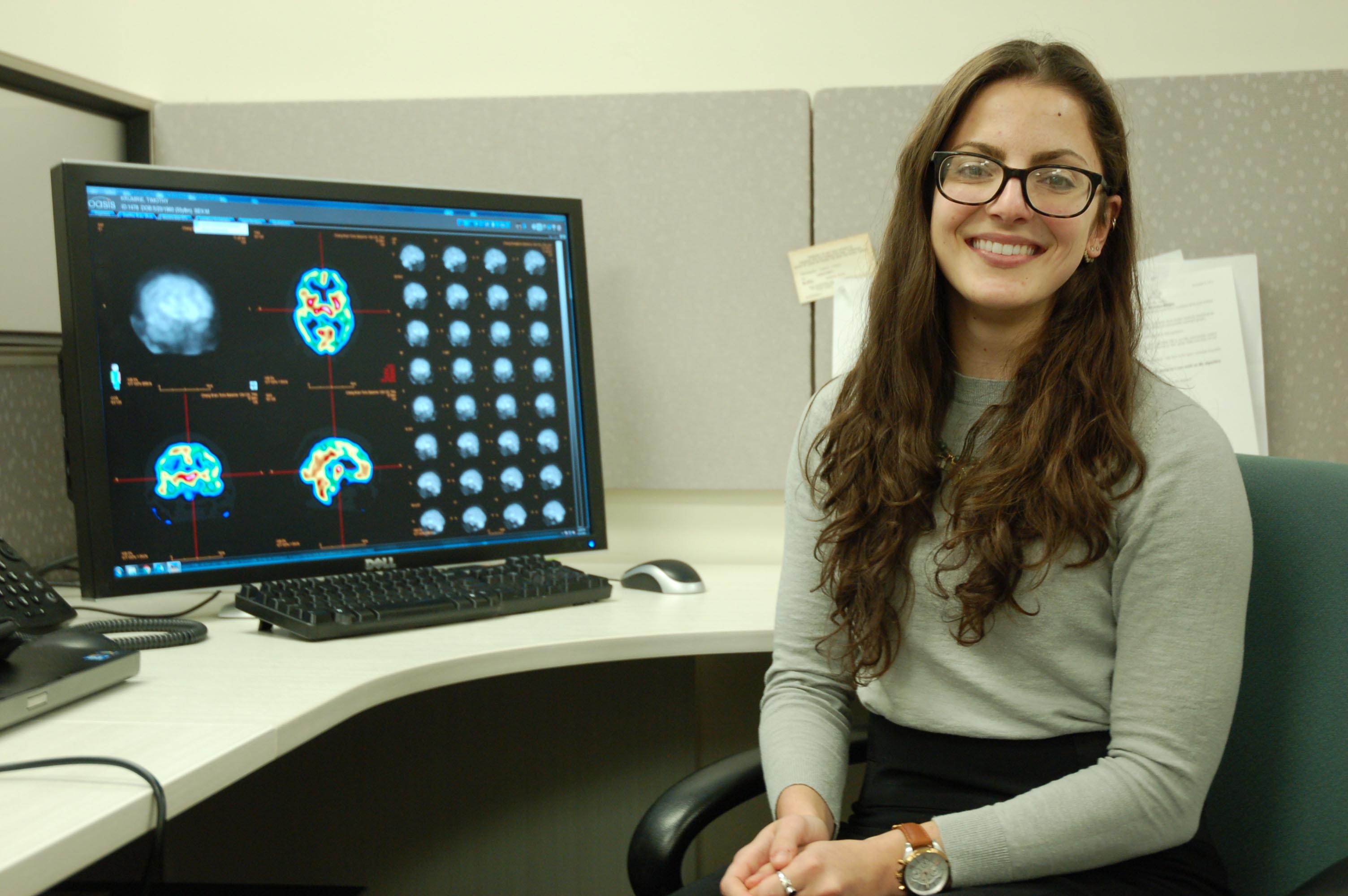
“We’re hoping to find patterns that will guide diagnosis,” said Lindsay Quandt, an imaging applications engineer at Cerescan.
Composed of clinicians, scientists, and technologists, the Cerescan team works together to ensure patients are scanned according to the company’s rigorous standards. Prior to imaging, each patient undergoes a thorough interview to provide clinicians with a comprehensive sense of their ailment.
Once a patient is scanned, the data generated by the scan is arranged into a matrix of voxels, which are tiny computer representations of clusters of cells. These voxels are generated from the gamma rays recorded by the imaging machine: the more blood flowing through each cluster of cells, the more gamma rays registered in the corresponding voxel.
The voxels produced by the imaging systems can be arranged to represent anatomical structures in a patient’s brain. By comparing the blood flowing through these anatomical structures to scans of healthy brains, Cerescan can identify the presence of disorders at a very fine level, leading in many cases to more precise diagnoses and more accurate measurements of treatment efficacy.
“At times, it can be a lot like CSI,” Kelley admitted, referring to CBS’s long-running detective show. “Sometimes it can get emotional, too. A father brought his son in recently. The son was suffering from addiction, and they were at wit’s end. The father, who was an acquaintance, had tried everything. He had even sent his son to camp in Idaho. As a final attempt at intervention, they came here.”
The doctor assigned to the case sat down with the son after reviewing his scans. “Son, your parents love you,” the doctor explained. “I’ve talked to them, and they just want you back.”
Cerescan’s technology allowed the doctor to determine that the son had not been taking his medication. It also allowed the doctor to identify an abnormally high amount of blood flow in the son’s occipital lobes.
“Then the doctor said, ‘When did you start hearing voices?’ Now, Cerescan doesn’t show voices, but the doctor had seen enough cases where the back of the brain was an issue.”
“And the son replied, ‘How did you know?”
Growing Recognition of the Technology’s Potential
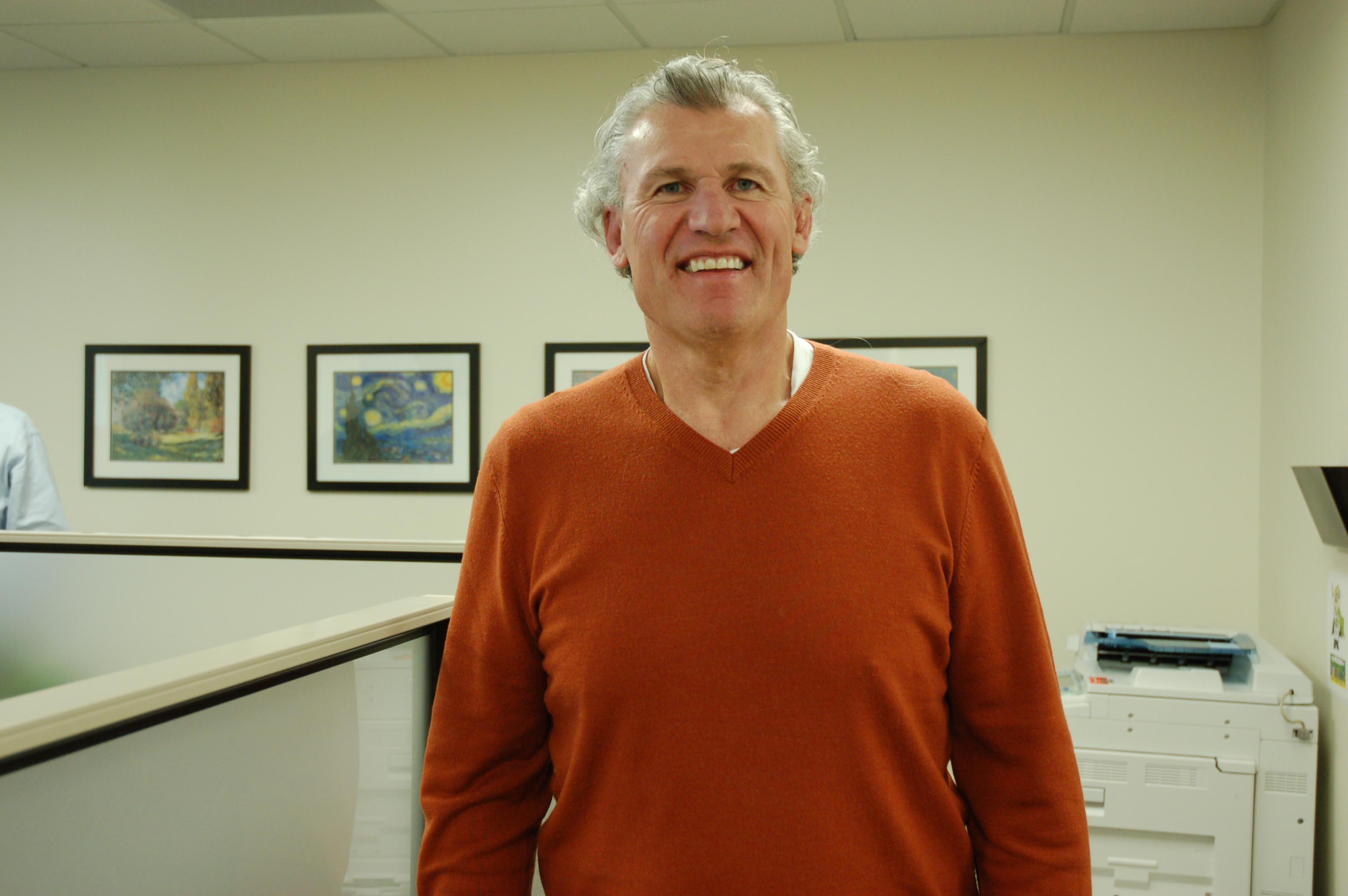
Former NFL player and coach Tim Krumrie works with Cerescan to help expand awareness of the company’s technology.
Cerescan is currently exploring the application of its technology to a variety of different disorders. Several studies are underway to test the technology’s ability to improve the diagnosis and treatment of conditions like concussion, depression, and attention deficit disorder (ADD).
“In December we were asked by a doctor how many of the patients we had scanned were suicidal,” explained Kelly. “It turned out that we had scanned 291 suicidal patients. One of our clinicians noticed that in two of the scans she had performed, there were three standard deviations of increased blood flow in the thalamic region. When we examined all 291 patients, we found that 141 of them had that same increase in blood flow.”
While Cerescan’s technology could be used anywhere that an imaging machine and a connection to the Internet are present, the company currently operates out of offices in Colorado, Texas, Florida, Kansas, and Louisiana. Because of the novelty of its technology, most of the patients Cerescan sees come to the imaging company only after having exhausted other options.
“We only see patients when the doctors say there’s a legitimate need,” admitted Kelley. “There has to be a qualified medical referral.”
Yet the company’s recent announcement that it had been awarded a quarter of a million dollars by the State of Colorado’s Office of Economic Development (OED) suggests that Cerescan’s brain imaging technology may soon be available to a far wider range of patients.
In a press release detailing the OED grant, the company stated that the funds would be used to commercialize its technology “for use by hospitals, clinics, physicians, and other healthcare practitioners as an advanced utility for diagnosis, treatment, and research into most neurological disorders.”
The Ability to See Improvement
“I played for the Cincinnati Bengals for twelve years,” Tim Krumrie, one of Cerescan’s brain injury advocates, told CyberMed News. “Obviously something was going to be off.”
After being introduced to the company by a friend, Krumrie scheduled a scan, which was followed by a treatment program that his doctor recommended based on Cerescan’s findings. For Krumrie, who recently spoke to the press about his experience with Cerescan during the build-up to Super Bowl 50, the results were dramatic.
“Stories came back. Long-term memories came back. But here’s the kicker. When I went back in and got rescanned, I looked at the before and after images, and I could actually see that my brain had improved.”
Like the coverage that CyberMed News provides? Follow us on Twitter, LinkedIn, and Facebook to make sure you keep up to date on the most recent developments in digital health.


Be the first to comment on "Harnessing the Big Data of Medical Imaging to Reveal the Brain’s Inner Workings"